
Learn more about the Basic Echo in Life Support Course
Objectives
On completion of this one-day clinically focused ultrasound program, emergency physicians will:- Use ultrasound system controls to optimise imaging for accurate diagnosis
- Develop proficiency in probe manipulation to efficiently acquire diagnostic images
- Describe the limitations and clinical integration of the basic echo in life support examination
- Use a systematic approach to conduct cardiac ultrasound assessment of a shocked or arrested patient
- Use ultrasound to assess the patient’s fluid status
CME recognition
|
|
|

CPD Short Format Learning |
|

CPD Categories 2B & 3B |
 Focused Echocardiography in Life Support
Features
- Modules (total) 3
- Skill level Novice
- Duration (days) 1
- Lectures (hours) 2
- Scan time (hours) 4.25
- Clinician Yes
- Zedu redo Yes
Class times
Day 1: 0830 – 1700
Modules
Image optimisation – interactive practical session
- Explore system controls
- Understand the impact of optimisation on the image
- Presets, frequency, depth, focus and gain
- How to label the image
- How to store and review images
Probe Manipulation
- Probe care
- Probe moves
- Probe grip
- Probe and screen orientation
- 3D awareness of the anatomical position of the organs
Basic Echo in Life Support
- anatomy of the heart in 3D – an interactive practical
- the 4 basic sonographic views of the heart
- LV and RV function
- Tamponade and PE
- Sepsis and hypovolaemia
- Volume status
- Limitations and pitfalls
- Techniques to improve imaging
- The role of cardiac ultrasound in advanced life support
- Clinical utility and application
- Case vignettes
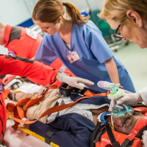
Scanning, scanning and more scanning of real people
CME details
ACEM | This workshop meets ACEM ultrasound guidelines and policies for ECHO. Approved for 8.5 ACEM CPD hours.
ACP | This course is recognised by the Australasian College of Paramedicine for CPD hours.
ANZCA | Participants in the ANZCA CPD program can claim attendance under the Knowledge and Skills category ‘Learning sessions’ at 1 credit per hour for the lectures/presentations. Hands on workshops may be claimed under the Knowledge and skills category ‘Short format learning’ at 2 credits per hour.
ASUM | CCPU Units – Focused Echocardiography in Life Support
CICM | This activity has CICM CPD Accreditation for Category 2B: Active of Interactive Small Group Learning – 2 points per hour | Category 3B: Quality Assurance and Patient Safety Activities – 3 points per hour.
A great training course is only the beginning of your ultrasound learning journey
To help you master ultrasound and lasting change from training we engage you from the outset
Before your course:
There are pre course activities & access to preparatory learning resources to maximise your in-class experience
During your course:
- Short, sharp information sessions to provide foundational knowledge and clinical context
- Lots of hands-on practical sessions with real people to scan (including for TV and obstetric scans). Our standardised patients models vary in age, size, shape and mobility to make the transition to clinical reality easier.
- Case studies led by practising clinicians to demonstrate clinical utility
- A variety of different machines to learn on
- At the conclusion of your course we’ll lead you to create a skills development action plan, creating a pathway that you can use to apply your newly acquired skills, promote clinical integration and ultimately reach your final objective – ultrasound competency
After your course:
The learning continues
- We encourage self-directed learning in the workplace with structured active learning plans
- A great selection of free access online medical ultrasound (FoamUS) learning resources
- You can subscribe to the weekly wrap – a weekly curation of ultrasound related hot tips and journal articles
- Be a part of our community of clever – Join our peer support network with our monthly “Coaching Corner” sessions. Send in your questions, join the live online meeting and have your questions answered.
The Zedu Redo – our commitment to you
We encourage you to refresh your training with us by taking up the opportunity to return within 12 months and do a complimentary redo of this course at no cost (subject to availability)
More Ways Zedu Can support your ultrasound skills development:
- Image review – details here
- Follow-up coaching with our Zedu coaches
- Coaching corner – monthly webinar
Basic Echo in Life Support Course
$1,250.00
At the completion of this course you will have developed a systematic approach to conduct cardiac ultrasound assessment of a shocked or arrested patient, and the ability to use ultrasound to assess the patient’s fluid status.
Select your date and enter your details to start your journey with us – we can’t wait to meet you.
Is your preferred course date full? Join our Waitlist Here

You may also like…
-
Focused Cardiac Ultrasound Course$3,465.00
Move beyond the basics and build your confidence to perform and interpret cardiac ultrasound. Master your spatial reasoning skills to get the best image possible for diagnosis.
Select your…
Select options- 3 days -
PE
Tamponade
RV/LV function
Doppler
Lung
Integration in arrest
-
Advanced Ultrasound for Emergency Medicine Course$4,550.00
Master advanced point of care ultrasound with this jam-packed course. Full of tips and tricks to help you make accurate diagnoses on the spot.
Select your date and enter your…
Select options- 4 days -
DVT
Biliary
Nerve Block
Early pregnancy
Soft tissue
Scrotum
Renal
Ocular
-
eFAST Ultrasound Course
Time poor or need to refresh? Want focused skills in a day? Learn how to perform an eFAST scan at our eFAST ultrasound course to…
Select options- 1 day -
eFAST
Run on demand


Related courses
-
MTOP Ultrasound Course$2,600.00
Confirm an IUP, ectopic pregnancy, RPOC, IUCD position, estimate gestational age, identify cause of pain & bleeding.
Select your date and enter your details to start your ultrasound…
Select options- 2 days -
Early pregnancy assessment
Pregnancy dating
Ectopic pregnancy
RPOC
IUCD placement
Live TA & TV scanning
-
Pet Ultrasound Course – FAST$0.00
Is your preferred course date full? Join our Waitlist Here
Select optionsImage optimsation
A-fast
T-fast
-
Ultrasound Guided Regional Anaesthesia Course$2,399.00
Use ultrasound to safely guide regional anaesthesia & reduce complications
Is your preferred course date full? Join our Waitlist Here
Select options- 2 days -
Image optimisation
Needle guidance techniques
Hip blocks
Knee blocks
Shoulder blocks
Truncal blocks


![]()
|
General Enquiries / Patient Models:
|
0401 810 585 |
| Clinical & training | 0422 000 750 |
|
Website / Registration:
|
0422 538 825 |
![]()
ZEDU Ultrasound Training Solutions
Suite 2, Level 1, 8-10 Flintoff Street
Greensborough VIC 3088 Australia